Restore Your Confidence: The Power Of Dental Bonding In Manassas Park
Dental bonding is a transformative solution that can address a variety of cosmetic dental concerns, helping individuals restore their smile and boost their confidence. Whether you have chipped, cracked, or discolored teeth, dental bonding offers a quick and effective way to enhance the appearance of your teeth with minimal discomfort and expense. In Manassas Park, this procedure is becoming increasingly popular for its versatility and natural-looking results. This article explores the benefits of dental bonding and how it can be a game-changer for those looking to improve their smile.
How To Know If You're A Good Candidate For Dental Bonding In Manassas Park
Dental bonding in Manassas Park is a great option for improving minor cosmetic issues like chips, cracks, or discoloration. It’s a non-invasive, affordable procedure that provides quick results, but it’s important to know if you're a good candidate before proceeding.
Minor Cosmetic Issues
Dental bonding is ideal for fixing small imperfections, like chips or stains. For larger issues, more advanced treatments may be needed.
Healthy Teeth And Gums
Candidates should have no active cavities or gum disease. Bonding works best on healthy teeth.
Good Oral Hygiene
Proper dental care is essential for the longevity of bonding. Regular brushing and flossing help maintain results.
Realistic Expectations
While dental bonding improves your smile, it may not be as durable as other treatments like crowns or veneers.
Non-Smokers Or Light Smokers
Smoking can stain bonding material. If you smoke, discuss this with your dentist to assess suitability.
Consulting a dentist in Manassas Park can help determine if dental bonding is the best option for your needs and set realistic expectations for your smile transformation.
What Are The Benefits That Can Come From Dental Bonding In Manassas Park
Dental bonding in Manassas Park offers several benefits, making it a popular choice for individuals looking to improve their smiles. Here are some of the key advantages.
- Dental bonding is a fast procedure that can often be completed in a single visit, without the need for invasive treatments like drills or surgery. It requires minimal preparation, making it an easy choice for those seeking a quick solution.
- Compared to other cosmetic treatments, such as veneers or crowns, dental bonding is more budget-friendly. It offers an effective way to address minor imperfections without breaking the bank.
- The resin used in dental bonding is color-matched to your natural teeth, ensuring a seamless, natural-looking result. This makes it an excellent option for addressing visible imperfections without affecting the overall appearance of your smile.
- Dental bonding can fix a variety of cosmetic issues, including chipped, cracked, or discolored teeth, as well as gaps between teeth. It can also be used to reshape or lengthen teeth for a more even and attractive smile.
- Unlike more invasive procedures, dental bonding typically causes little to no discomfort. Most patients experience only mild sensitivity, which is temporary and fades shortly after the procedure.
- Since bonding is completed in a single visit, patients can walk out with a restored smile right away. This immediate transformation is a major benefit for those seeking quick cosmetic improvements.
Overall, dental bonding in Manassas Park is a fast, affordable, and effective way to enhance your smile with minimal disruption to your daily routine. For the best results, it's important to choose a reputable dentist with experience in cosmetic procedures, such as the skilled professionals at Railroad Dental Associates. Their expertise ensures that you receive personalized care, high-quality materials, and results that not only improve the appearance of your teeth but also boost your confidence.
How To Choose A Dentist In Manassas Park, VA, That Specializes In Dental Bonding
Choosing a dentist in Manassas Park, VA, who specializes in dental bonding involves considering their experience, expertise, and patient care approach. Start by researching local dentists who offer cosmetic dentistry services, particularly those with a strong track record in performing dental bonding procedures.
Look for a dentist who has experience with a variety of cosmetic treatments and uses up-to-date materials and techniques. This ensures that you receive the best possible care and results for your specific needs.
Patient reviews and testimonials are also valuable; they provide insights into the quality of care, the dentist’s skill in achieving natural-looking results, and overall satisfaction. This feedback can help you gauge whether the dentist’s approach aligns with your expectations.
During your consultation, ensure the dentist takes the time to understand your goals and explain the process clearly, while also assessing your specific dental needs. A good dentist will offer personalized recommendations, provide realistic expectations, and create a comfortable environment for the procedure.
What To Expect During A Dental Bonding Procedure In Manassas Park
During a dental bonding procedure in Manassas Park, you can expect a straightforward and relatively comfortable experience. Here's a breakdown of what to anticipate.
Consultation And Preparation
Before the procedure begins, your dentist will assess the areas you want to improve. They may discuss your cosmetic goals and decide whether dental bonding is the best option. The affected teeth will be cleaned, and a local anesthetic may be used if necessary, though bonding is usually painless and doesn’t require numbing.
Resin Application
The dentist will apply a tooth-colored resin to the area being treated. The resin is soft and pliable, allowing the dentist to mold it into the desired shape to fix chips, cracks, or other imperfections. The dentist will ensure that the resin matches the natural color of your teeth for a seamless appearance.
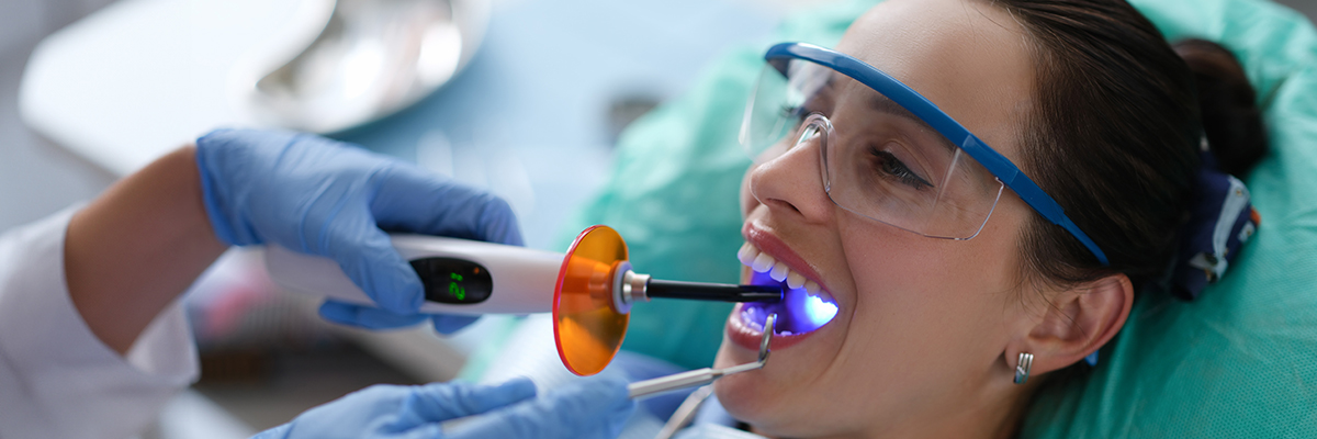
Hardening With A Special Light
Once the resin is shaped, a special light is used to harden and bond the material to your tooth. This typically takes just a few minutes and ensures that the resin stays securely in place.
Shaping And Polishing
After the resin has set, your dentist will trim and shape it to ensure it fits naturally with your other teeth. They will also polish the bonded area, making it smooth and giving it a shiny, natural appearance.
Completion
The procedure is quick, usually taking between 30 minutes to an hour, depending on the number of teeth being treated. You’ll leave the office with a restored smile, and there’s typically no recovery time required.
Overall, dental bonding is a fast, non-invasive procedure with minimal discomfort, providing immediate results that can significantly improve the appearance of your teeth.
What Are Some Tips For Ensuring The Longevity Of Your Dental Bonding In Manassas Park
To ensure the longevity of your dental bonding in Manassas Park, it’s important to follow a few key tips to maintain the results and prevent damage. Here are some helpful guidelines.
- Regular brushing and flossing are essential for maintaining both your natural teeth and bonded areas. Be sure to use a soft-bristled toothbrush and non-abrasive toothpaste to avoid scratching the bonding material.
- While dental bonding is durable, it’s still important to avoid biting down on hard objects like ice, pens, or hard candy, which can chip or crack the bonding material. Sticky foods like caramel or chewing gum can also pull at the bond, potentially causing it to wear down faster.
- Although bonding resin is color-matched to your teeth, it can still absorb stains over time. Limiting your intake of coffee, tea, red wine, and other staining substances will help keep your bonding looking fresh and bright.
- Smoking can stain both your natural teeth and the bonding material, reducing the aesthetic appeal of the procedure. If you smoke, it’s a good idea to reduce or quit to maintain the results for as long as possible.
- If you play sports or grind your teeth at night (bruxism), wearing a mouthguard can protect your dental bonding from damage. Grinding can cause the bonding material to wear down prematurely, so a mouthguard is an effective preventive measure.
- Routine dental visits are important for monitoring the condition of your bonding and ensuring your overall oral health. Your dentist can check for any signs of wear and tear and make adjustments or repairs as needed.
By following these simple tips, you can ensure that your dental bonding in Manassas Park, VA, lasts as long as possible, maintaining a beautiful, functional smile for years to come.
Contact A Dentist In Manassas Park, VA
Dental bonding in Manassas Park offers a transformative, affordable, and effective solution for restoring your smile and boosting your confidence. Whether you’re looking to fix chipped, cracked, or discolored teeth, bonding provides a quick, non-invasive treatment that delivers natural-looking results.
If you're considering dental bonding to restore your smile, Railroad Dental Associates in Manassas Park is there to help. With their expertise in cosmetic dentistry, they can assess your needs and guide you through the process, ensuring that dental bonding is the right choice for you. Contact them to learn more.